

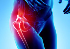
Outpatient Hip Replacement
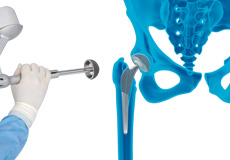
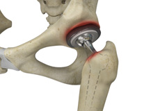
Hip replacement surgery is one of the most common orthopedic surgeries performed. It involves the replacement of the damaged hip bone (ball shaped upper end of the femur) with a ceramic ball attached to a metal stem that is fixed into the femur and placing a new cup with a special liner in the pelvis.
Robotic Hip Replacement
Mako Robotic-Arm Assisted Total Hip replacement is a surgical procedure intended for patients who suffer from non-inflammatory or inflammatory degenerative joint disease (DJD).
Muscle Sparing Anterior Hip Replacement
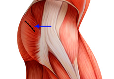
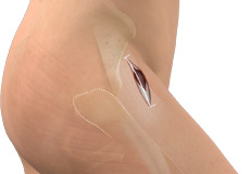
Muscle sparing anterior hip replacement is a minimally invasive hip surgery to replace the hip joint without cutting through any muscles or tendons as compared to traditional hip replacement that involves cutting major muscles to access the hip joint.
Mini-Posterior Hip Replacement
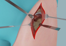
Mini-posterior hip replacement is a surgical procedure used to replace your damaged hip with synthetic parts inserted through a small incision made at the back of the hip.
Revision Hip Replacement
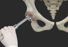
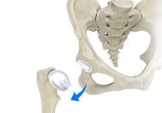
During total hip replacement, the damaged cartilage and bone are removed from the hip joint and replaced with artificial components.
Revision Hip Surgery
10-20% of people continue to have pain after joint replacement. Pain can be related to several reasons including wear, loosening, malposition of the implants, fracture and infection.
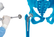
Robotic Total Hip Replacement
Robotic total hip replacement is a minimally invasive procedure where your surgeon is assisted by a robotic system to perform a total hip replacement surgery.
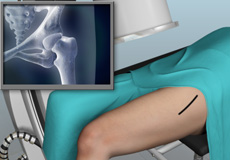
Outpatient Anterior Approach Hip Replacement
With improved technology and advances in anesthesia and pain control, hip replacement surgery has evolved and is now being offered in an outpatient setting.

Activities After Hip Replacement
You will be discharged from the hospital once you have sufficient pain control and are able to perform basic activities on your own, such as getting in and out of bed, going to the bathroom and walking with an assistive device such as crutches or a walker.
BMAC Of the Hip
BMAC is a concentrate of regenerative stem cells obtained from your own bone marrow.
Minimally Invasive Anterior Hip Replacement with X-Ray Guidance
Direct anterior hip replacement is a minimally invasive hip surgery to replace the hip joint without cutting through any muscles or tendons as against traditional hip replacement that involves cutting major muscles to access the hip joint.
Robotic Assisted Hip Surgery
Robotic assisted hip surgery is a minimally invasive surgical procedure that involves the use of a specialized robotic system to remove the damaged parts of a hip joint and replace them with an artificial prosthesis or implant.
Joint Replacement Infection
A very small percentage of patients (less than 1%) who undergo hip replacement may develop an infection around the hip joint following surgery. This infection is called a periprosthetic hip infection.
Physical Examination of the Hip
The physical examination of the hip by your doctor includes a visual inspection of your hip, palpation of the hip to diagnose tenderness or any abnormality, etc; and testing range of motion of the hip.
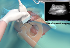
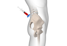
Ultrasound-Guided Hip Injections
An ultrasound scan is an imaging procedure that uses high-frequency sound waves to produce pictures of the inside of the body.
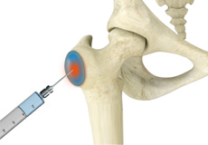
Trochanteric Bursa Injection
A trochanteric bursa injection is a minimally invasive procedure in which medicine is injected directly into the trochanteric bursa in the hip joint using a thin needle and syringe to relieve pain and inflammation.
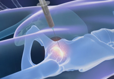
Stem Cell Therapy for Hip Injuries
Stem cell therapy is a form of regenerative medicine that utilizes the body’s natural healing mechanism to treat various conditions.
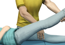
Physical Therapy for Hip
Physical therapy is an exercise program that helps you to improve movement, relieve pain, encourage blood flow for faster healing, and restore your physical function and fitness level.
Iliopsoas Tendonitis
Iliopsoas tendonitis also referred to as snapping hip syndrome, is an inflammation of the iliopsoas tendon or the surrounding area.
Stress Fractures of the Hip
Stress fractures of the hip are a break in the upper part of the thigh bone (femur) that fits into the socket of the hip joint.
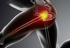
Hip Injury
The hip joint is one of the most important and flexible joints in the human body which allows us to walk, run, bend and perform physical activities.
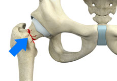
Femoral Neck Fracture
The hip is a ball-and-socket joint made up of the head of the thigh bone or femur that acts as the ball and fits into the rounded socket of the hip bone or acetabulum.
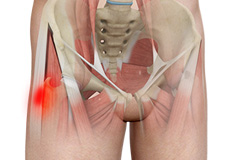
Hip Bursitis
Hip bursitis is a painful condition caused by the inflammation of a bursa in the hip. Bursae are fluid-filled sacs present in the joints between bone and soft tissue to reduce friction and provide cushioning during movement.
Hip Fracture
The hip joint is a “ball and socket” joint. The “ball” is the head of the femur or thighbone, and the “socket” is the cup-shaped acetabulum.
Hip Dislocation
The hip joint is a “ball and socket” joint. The “ball” is the head of the femur or thighbone, and the “socket” is the cup-shaped acetabulum.
Hip Tendonitis
Tendons are strong connective tissue structures that connect muscle to bone. Hip tendonitis is a condition associated with degeneration of the hip tendons.
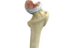
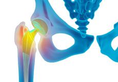
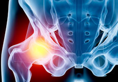
Osteoarthritis of the Hip
Osteoarthritis, also called degenerative joint disease, is the most common form of arthritis. It occurs most often in the elderly.
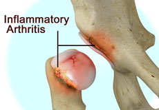
Inflammatory Arthritis of the Hip
The inflammation of the joints is referred to as arthritis. Inflammation arises when the smooth lining called cartilage at the ends of bones wears away.
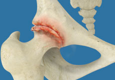
Hip Osteonecrosis
Hip osteonecrosis occurs due to disruption of the blood supply to the highest part of the thigh bone (femoral head).
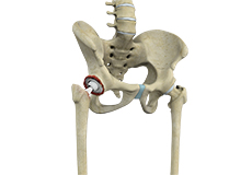
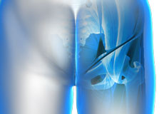
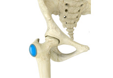
Hip Joint
The hip joint is the largest weight-bearing joint in the human body. It is also referred to as a ball and socket joint and is surrounded by muscles, ligaments, and tendons. The thigh bone or femur and the pelvis join to form the hip joint.
Any injury or disease of the hip will adversely affect the joint's range of motion and ability to bear weight.
The hip joint is made up of the following:
- Bones and joints
- Ligaments of the joint capsule
- Muscles and tendons
- Nerves and blood vessels that supply the bones and muscles of the hip
Bones and Joints
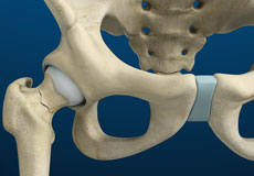
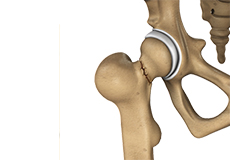
The hip joint is the junction where the hip joins the leg to the trunk of the body. It is comprised of two bones: the thigh bone or femur and the pelvis which is made up of three bones called ilium, ischium, and pubis. The ball of the hip joint is made by the femoral head while the socket is formed by the acetabulum. The Acetabulum is a deep, circular socket formed on the outer edge of the pelvis by the union of three bones: ilium, ischium, and pubis. The lower part of the ilium is attached by the pubis while the ischium is considerably behind the pubis. The stability of the hip is provided by the joint capsule or acetabulum and the muscles and ligaments which surround and support the hip joint.
The head of the femur rotates and glides within the acetabulum. A fibrocartilaginous lining called the labrum is attached to the acetabulum and further increases the depth of the socket.
The femur or thigh bone is one of the longest bones in the human body. The upper part of the thigh bone consists of the femoral head, femoral neck, and greater and lesser trochanters. The head of the femur joins the pelvis (acetabulum) to form the hip joint. Next, to the femoral neck, there are two protrusions known as greater and lesser trochanters which serve as sites of muscle attachment.
Articular cartilage is the thin, tough, flexible, and slippery surface lubricated by synovial fluid that covers the weight-bearing bones of the body. It enables smooth movements of the bones and reduces friction.
Ligaments
Ligaments are fibrous structures that connect bones to other bones. The hip joint is encircled with ligaments to provide stability to the hip by forming a dense and fibrous structure around the joint capsule. The ligaments adjoining the hip joint include:
- Iliofemoral ligament: This is a Y-shaped ligament that connects the pelvis to the femoral head at the front of the joint. It helps in limiting the over-extension of the hip.
- Pubofemoral ligament: This is a triangular shaped ligament that extends between the upper portion of the pubis and the iliofemoral ligament. It attaches the pubis to the femoral head.
- Ischiofemoral ligament: This is a group of strong fibers that arise from the ischium behind the acetabulum and merge with the fibers of the joint capsule.
- Ligamentum teres: This is a small ligament that extends from the tip of the femoral head to the acetabulum. Although it has no role in hip movement, it does have a small artery within that supplies blood to a part of the femoral head.
- Acetabular labrum: The labrum is a fibrous cartilage ring which lines the acetabular socket. It deepens the cavity, increasing the stability and strength of the hip joint.
Muscles and Tendons
A long tendon called the iliotibial band runs along the femur from the hip to the knee and serves as an attachment site for several hip muscles including the following:
- Gluteal: These are the muscles that form the buttocks. There are three muscles (gluteus minimus, gluteus maximus, and gluteus medius) that attach to the back of the pelvis and insert into the greater trochanter of the femur.
- Adductors: These muscles are located in the thigh which helps in adduction, the action of pulling the leg back towards the midline.
- Iliopsoas: This muscle is located in front of the hip joint and provides flexion. It is a deep muscle that originates from the lower back and pelvis and extends up to the inside surface of the upper part of the femur.
- Rectus femoris: This is the largest band of muscles located in front of the thigh. They also are hip flexors.
- Hamstring muscles: These begin at the bottom of the pelvis and run down the back of the thigh. Because they cross the back of the hip joint, they help in extension of the hip by pulling it backward.
Nerves and Arteries
Nerves of the hip transfer signals from the brain to the muscles to aid in hip movement. They also carry the sensory signals such as touch, pain, and temperature back to the brain.
The main nerves in the hip region include the femoral nerve in the front of the femur and the sciatic nerve at the back. The hip is also supplied by a smaller nerve known as the obturator nerve.
In addition to these nerves, there are blood vessels that supply blood to the lower limbs. The femoral artery, one of the largest arteries in the body, arises deep in the pelvis and can be felt in front of the upper thigh.
Hip Movements
All of the anatomical parts of the hip work together to enable various hip movements. Hip movements include flexion, extension, abduction, adduction, circumduction, and hip rotation.